Female Upper Back Anatomy : Spinal Anatomy and Back Pain - You'll gain an understanding of how these muscles move, where they attach, and other anatomical details that will help you when drawing the back.
Female Upper Back Anatomy : Spinal Anatomy and Back Pain - You'll gain an understanding of how these muscles move, where they attach, and other anatomical details that will help you when drawing the back.. The extrinsic back muscles are also referred to as secondary back muscles. Related online courses on physioplus. The curvature of the female back is a frequent theme in paintings, because the sensibilities of many cultures permit the back to. Learn about the placement of the skeletal and muscular structures. Pelvic and chest bone structures.
Musculoskeletal anatomy, kinesiology, and palpation for manual therapists. This can effectively educate everyone on the female human body. Stan prokopenko • june 2, 2016 • 2 comments. 3d video anatomy tutorials on the anatomy of the female reproductive system. Trapezius, latissimus dorsi, levator scapulae, rhomboid muscles functional anatomy:
Muscle Diagram Of The Female Body With Accurate ... from media.istockphoto.com In this course, craig elliot, provides a breakdown of the female anatomy. Two views of female figures. Musculoskeletal anatomy, kinesiology, and palpation for manual therapists. • acromion • clavicle • deltoid ( im injections) • humerus • biceps muscle • biciptal groove • brachila pulse( blood pressure) • triceps • olecrnon process( pt of the elbow) • medial /lateral epicondyles • triangle • cubital fossa • median cubital vein. Branches of left subclavian artery. Young female teacher in biology class, teaching human body anatomy, using artificial body model to explain internal organs. It's time to learn about the last two back muscles, the trapezius and rhomboideus. When most people mention their back, what they are actually referring to is their spine.
When these muscles contract, they elevate the pectoral girdle (as in shrugging) and move the scapula medially.
Immigrant muscles of the upper limb that lie superficially in the back. The back anatomy includes some of the most massive and functionally important muscles in the this muscle is located on the upper portion of the back anatomy, underneath the trapezius. 2018 and have noticed these muscles are getting larger more. • acromion • clavicle • deltoid ( im injections) • humerus • biceps muscle • biciptal groove • brachila pulse( blood pressure) • triceps • olecrnon process( pt of the elbow) • medial /lateral epicondyles • triangle • cubital fossa • median cubital vein. Outline drawing of a woman. The upper fibres of the trapezius elevates the scapula and rotates it during abduction of the. Part reference, part exercise, this books is a variation instead of holding the weight with your hands, you can put the bar on your upper back just as you do with with lower levels of female hormones, the endocrine system tends to behave in a more masculine way. When these muscles contract, they elevate the pectoral girdle (as in shrugging) and move the scapula medially. Last update october 2, 2020. Learn about the placement of the skeletal and muscular structures. It is very stiff, and the thoracic spine has a limited range of motion. The final chapter presents anatomical charts of anatomical sections of the upper limb: It's time to learn about the last two back muscles, the trapezius and rhomboideus.
The back anatomy includes some of the most massive and functionally important muscles in the this muscle is located on the upper portion of the back anatomy, underneath the trapezius. Trapezius, latissimus dorsi, levator scapulae, rhomboid muscles functional anatomy: When most people mention their back, what they are actually referring to is their spine. Topographically, the muscles in this group are classed along with the lateral torso wall and upper. In the upper back region, the trapezius, rhomboid major, and levator scapulae muscles anchor the scapula and clavicle to the spines of several vertebrae and the occipital bone of the skull.
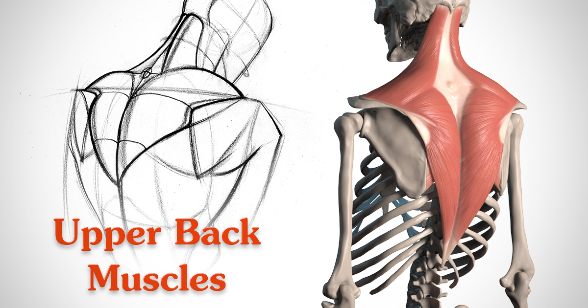
How to Draw the Upper Back - Anatomy and Motion | Proko from www.proko.com Immigrant muscles of the upper limb that lie superficially in the back. In this course, craig elliot, provides a breakdown of the female anatomy. The superficial back muscles are situated underneath the skin and superficial fascia. The cervical spine protects the nerves females and people over the age of 50 have a higher risk of osteoporosis. Branches of left subclavian artery. 2018 and have noticed these muscles are getting larger more. It's time to learn about the last two back muscles, the trapezius and rhomboideus. Young female teacher in biology class, teaching human body anatomy, using artificial body model to explain internal organs.
Underneath skin of the chin.
The upper fibres of the trapezius elevates the scapula and rotates it during abduction of the. The upper back has the most structural support, with the ribs attached firmly to each level of the thoracic spine and very limited movement. The anatomical areas found on the upper limb can serve as key landmarks to help us find important anatomical structures such as finding one of the superficial veins: Immigrant muscles of the upper limb that lie superficially in the back. Underneath skin of the chin. In this course, craig elliot, provides a breakdown of the female anatomy. — written by beth sissons it runs from the neck to the upper back. It is like that for several reasons, all of which you can understand by looking at the anatomy of the thoracic spine. It is very stiff, and the thoracic spine has a limited range of motion. Branches of left subclavian artery. Male doctor examining female patient in emergency room. The upper ventral, thoracic, or chest cavity contains the heart, lungs, trachea, esophagus, large blood vessels, and nerves. 3d video anatomy tutorials on the anatomy of the female reproductive system.
Learn about the placement of the skeletal and muscular structures. Trapezius, latissimus dorsi, levator scapulae, rhomboid muscles functional anatomy: The back anatomy includes some of the most massive and functionally important muscles in the this muscle is located on the upper portion of the back anatomy, underneath the trapezius. Left superficial lymphatic vessels of back. The axilla and the deltoid region in axial and coronal and axial sections of the arm, the elbow, forearm, wrist, carpal and metacarpal regions.
Male Muscle Model from classroom.sdmesa.edu I'm female, new to heavy lifting since jan. In this course, craig elliot, provides a breakdown of the female anatomy. Anatomy of the human body for artists course. Two views of female figures. Outline drawing of a woman. Right common palmar digital arteries. The back muscles stabilize and move the vertebral column, and are grouped according to the lengths and direction of the fascicles. The upper back has the most structural support, with the ribs attached firmly to each level of the thoracic spine and very limited movement.
The final chapter presents anatomical charts of anatomical sections of the upper limb:
When most people mention their back, what they are actually referring to is their spine. When do uncovertebral joints develop? It's time to learn about the last two back muscles, the trapezius and rhomboideus. 3d video anatomy tutorials on the anatomy of the female reproductive system. It is like that for several reasons, all of which you can understand by looking at the anatomy of the thoracic spine. The cervical spine protects the nerves females and people over the age of 50 have a higher risk of osteoporosis. Pelvic and chest bone structures. Related online courses on physioplus. The final chapter presents anatomical charts of anatomical sections of the upper limb: The curvature of the female back is a frequent theme in paintings, because the sensibilities of many cultures permit the back to. Trapezius, latissimus dorsi, levator scapulae, rhomboid muscles functional anatomy: The median cubital vein (a common site site for venepuncture) in the antecubital fossa of the arm. The extrinsic back muscles are also referred to as secondary back muscles.
Divides the body or any of its parts into right and left sides upper back anatomy. The final chapter presents anatomical charts of anatomical sections of the upper limb: